REVIEW ARTICLE
CARDIOVASCULAR
COMPLICATIONS IN ADULTS AFTER SARS-COV-2 INFECTION: A COMPARATIVE SYSTEMATIC
REVIEW
CARDIOVASCULAR
COMPLICATIONS IN ADULTS FOLLOWING SARS-CoV-2 INFECTION: A COMPARATIVE
SYSTEMATIC REVIEW
CARDIOVASCULAR
COMPLICATIONS IN ADULTS FOLLOWING SARS-COV-2 INFECTION: A COMPARATIVE
SYSTEMATIC REVIEW
https://doi.org/10.31011/reaid-2026-v.100-n.3-art.2685
1Vitória
Pereira Silva Dutra
2Solon
Alves da Silva Filho
3Antonio
Frivaldo Marinho Neto
4Marcos
Vinícius Novais Alves
5Rafael
Aires Oliveira Campos
6Karla
Vanessa Morais Lima
7Arlane
Silva Chaves
8Francisco
Alves Lima Júnior
1Universiadade
Federal do Maranhão, Imperatriz, Brazil. Orcid: 0000-0002-7636-3874
2Universidade
Ceuma, Imperatriz, Brazil. Orcid: 0009-0008-1463-5599
3Universidade
Ceuma, Imperatriz, Brazil. Orcid:0009-0003-7055-0200
4Unisulma,
Imperatriz, Brazil. Orcid: 0009-0009-5182-8995
5
Universidade Ceuma, Imperatriz, Brasil. Orcid: 0009-0000-9415-7217
6Faculdade
de Medicina de Açailândia, Açailãncia, Brazil. Orcid: 0000-0002-3018-2621
7Universiadade
Federal do Maranhão, Imperatriz, Brazil. Orcid: 0000-0002-6271-6237
8Universiadade
Federal do Maranhão, Imperatriz, Brazil. Orcid: 0000-0002-3117-4949
Corresponding
Author
Francisco
Alves Lima Júnior
Av.
Pedro Neiva de Santana, Ecopark III, casa 62. Bairro João - Paulo II,
Imperatriz, Maranhão, Brazil. CEP: 65.919-555. E-mail: francsico.alj@ufma.br
Submission: 05-11-2025
Approval: 07-11-2025
ABSTRACT
Objective: To identify cardiovascular complications
triggered by SARS-CoV-2 infection in patients with and without pre-existing
cardiovascular disease and to present the associated pathophysiological
mechanisms. Methods: Systematic literature review with
meta-analysis. Searches were performed in the PubMed, Web of Science, Lilacs,
and Scopus (Elsevier) databases. The following DeCS terms were used:
“SARS-CoV-2,” “COVID-19,” “cardiovascular diseases,” “post-COVID syndrome,” and
“cardiovascular abnormalities.” A total of 10 studies were included. Results:
Echocardiographic abnormalities were found to occur with similar probability
among all patients. Heart rate turbulence and variability remained abnormal
even six months after the acute phase, indicating cardiac autonomic dysfunction
and risk for further morbid cardiac complications. Alterations in left and
right ventricular diastolic function and a higher risk of venous thromboembolic
events were observed in patients with COVID-19. Endothelial dysfunction was
prevalent, especially in those with heart failure and acute myocardial injury.
Autonomic dysfunction may result from diffuse endotheliitis and vascular
injury. Ischemic myocardial injury arises from cytokine storm, catecholamine
surge, plaque rupture or erosion, hypoxic damage, coronary artery spasm, and
thrombosis. Heart failure is hypothesized to occur due to immune system
hyperactivation and downregulation of ACE2. Conclusion:
SARS-CoV-2 infection affects the cardiovascular system, leading to myocardial
injury, endothelial dysfunction, impaired myocardial oxygen supply and demand,
and global dysfunction of both left and right ventricles.
Keywords: SARS-CoV-2; COVID-19; Cardiovascular
Diseases; Post-COVID Syndrome; Cardiovascular Abnormalities.
RESUMO
Objetivo: Identificar as
complicações cardiovasculares desencadeadas pela infecção do Sars-CoV-2, em
pacientes com e sem Doenças Cardiovasculares e apresentar os mecanismos
fisiopatológicos. Métodos: Revisão sistemática da literatura com
metanálise. As buscas foram realizadas nas bases de dados: PubMed; Web of
Science; Lilacs e Scopus (Elsevier). Foi utilizado os DeCS: “sars-cov-2”,
“covid-19”, “doenças cardiovasculares”, “síndrome pós-covid”, “anormalidade
cardiovasculares. Seleção final de 10 estudos. Resultados: É observado
que anormalidades ecocardiográficas possuem a mesma probabilidade de ocorrer em
todos os pacientes, a turbulência da frequência cardíaca e a variabilidade da
frequência cardíaca foi anormal mesmo 6 meses após a fase aguda da doença,
indicando disfunção autonômica cardíaca e risco para o desenvolvimento de
outras complicações cardíacas mórbidas. Alterações na função diastólica do
ventrículo esquerdo e direito e o risco de desenvolvimento de eventos
tromboembólicos venosos em pacientes críticos é maior na presença de COVID-19.
Há uma prevalência de disfunção endotelial, principalmente em pacientes com insuficiência
cardíaca e lesão miocárdica aguda. Um dos mecanismos responsáveis
pela disfunção autonômica é a endotelite difusa e a lesão vascular. A lesão
miocárdica isquêmica resulta da tempestade de citocinas, picos de
catecolaminas, ruptura ou erosão da placa e lesão hipóxica, espasmo da artéria
coronária e trombose, postula-se que a insuficiência cardíaca ocorre devido à
hiperativação do sistema imunológico e à regulação negativa da ECA2. Conclusão: A infecção pelo vírus SARS-CoV-2 afeta o sistema CV
levando a lesão miocárdica, disfunção endotelial, disfunção na oferta e demanda
de oxigênio do miocárdio e disfunção global do ventrículo esquerdo e direito.
Palavras-chave:
Sars-cov-2; Covid-19; Doenças Cardiovasculares;
Síndrome pós-Covid; Anormalidade Cardiovasculares.
RESUMÉN
Objetivo: Identificar las complicaciones
cardiovasculares desencadenadas por la infección del SARS-CoV-2 en pacientes
con y sin enfermedades cardiovasculares previas y presentar los mecanismos
fisiopatológicos asociados. Métodos: Revisión sistemática de
la literatura con metanálisis. Las búsquedas se realizaron en las bases de
datos PubMed, Web of Science, Lilacs y Scopus (Elsevier). Se utilizaron los
DeCS: “SARS-CoV-2”, “COVID-19”, “enfermedades cardiovasculares”, “síndrome pos-COVID”
y “anormalidades cardiovasculares”. Se seleccionaron 10 estudios. Resultados:
Se observó que las anormalidades ecocardiográficas presentaron una probabilidad
similar de ocurrir en todos los pacientes. La turbulencia y la variabilidad de
la frecuencia cardíaca permanecieron alteradas incluso seis meses después de la
fase aguda, lo que indica disfunción autonómica cardíaca y riesgo de
complicaciones cardíacas mórbidas adicionales. Se identificaron alteraciones en
la función diastólica de los ventrículos izquierdo y derecho, y un mayor riesgo
de eventos tromboembólicos venosos en pacientes con COVID-19. La disfunción
endotelial fue prevalente, especialmente en casos con insuficiencia cardíaca y
lesión miocárdica aguda. La disfunción autonómica puede deberse a endotelitis
difusa y lesión vascular. La lesión miocárdica isquémica resulta de la tormenta
de citocinas, el aumento de catecolaminas, la ruptura o erosión de la placa, la
hipoxia, el espasmo coronario y la trombosis. Se postula que la insuficiencia
cardíaca ocurre por la hiperactivación del sistema inmunológico y la regulación
negativa de la ECA2. Conclusión: La infección por el virus
SARS-CoV-2 afecta el sistema cardiovascular, provocando lesión miocárdica,
disfunción endotelial, alteraciones en la oferta y demanda de oxígeno del
miocardio y disfunción global de los ventrículos izquierdo y derecho.
Palabras clave: SARS-CoV-2; COVID-19; Enfermedades
Cardiovasculares; Síndrome Pos-COVID; Anormalidades Cardiovasculares.
INTRODUCTION
COVID-19
is an infectious disease caused by the SARS-CoV-2 virus, which produces a wide
range of symptoms, varying from a common cold to severe respiratory
complications and systemic effects; there may even be asymptomatic cases1.
The
SARS-CoV-2 virus belongs to the Coronaviridae family; the best-known members of
this group are the Severe Acute Respiratory Syndrome Coronavirus (SARS-CoV) and
the Middle East Respiratory Syndrome Coronavirus (MERS-CoV). During 2002 and
2003, SARS-CoV was responsible for an outbreak of severe respiratory disease in
China, and MERS-CoV, in 2012, for an outbreak of severe respiratory diseases
centered in the Middle East2
On
January 31, 2019, the World Health Organization (WHO) received an alert from the
city of Wuhan, China, regarding severe pneumonia caused by a virus that, until
then, had no known history of infecting humans. This virus, now known as
SARS-CoV-2, quickly spread throughout the country and to other continents, and
was classified on January 30, 2020, as a Public Health Emergency of
International Concern (PHEIC) and subsequently, on March 11 of the same year,
as a pandemic, which did not end until three years later, on May 5, 2023 3.
The
coronavirus pandemic, as it became known, had recorded, as of May 26, 2024,
approximately 775 million confirmed cases of the disease and 7 million deaths
worldwide 4. According to the Coronavirus Dashboard, in Brazil, as
of July 8, 2024, there were 38,832,913 confirmed cases and 712,537 reported
deaths 5.
The
SARS-CoV-2 virus is an enveloped virus containing an RNA genome; there are 4
groups and 7 strains that infect humans. According to Sharma, studies reveal
that the SARS-CoV-2 genome is 96.2% similar to the genome of SARS RaTG13, which
very commonly infects bats—compared to SARS-CoV and MERS-CoV, which share 79%
and 51.8% similarity, respectively. Thus, it is suggested that bats are the
origin of SARS-CoV-2 6.
According
to Rodrigues 7, the SARS-CoV-2 virus enters the cell via the
angiotensinogen-converting enzyme (ACE2), which is found in the alveolar
epithelium, as well as in renal, cardiac, and gastrointestinal cells and blood
vessels, and plays a key role in the renin-angiotensin-aldosterone system by regulating
blood pressure and fluid volume within the vessels. Because its cellular
receptor is an enzyme present in so many organs of the body and due to the
body’s own defense against the pathogen, the pathophysiology of COVID-19 is
complex and is not restricted to a single system.
In
this regard, in the lungs, the first organ affected because they serve as the
entry point—the virus induces inflammatory and immune reactions, primarily in
the interstitial tissue of the lungs and the alveoli, leading to exudate
accumulation, and lesions in the alveolar and vascular epithelium trigger an
inflammatory cascade and consequent acute respiratory distress, while the
destruction of type II pneumocytes leads to atelectasis due to a lack of
surfactant 7.
The
virus has been detected in renal tissue, causing acute kidney injury,
proteinuria, and edema. In the liver, the cytopathic effect of the virus and
the immune response lead to sepsis and liver injury; in the enterocytes of the
small intestine, it causes intestinal dysbiosis, reducing immunity. In turn, in
the central and peripheral nervous systems, it induces headache, anosmia, and
hyposmia, and may lead to intracerebral and subarachnoid hemorrhages.
Furthermore, it can trigger cytokine production, leading to disseminated
intravascular coagulation (DIC), thrombus formation, and vascular occlusion 8.
Because
the virus uses the ACE2 enzyme, which is directly linked to the cardiovascular
system, as its cellular entry point, significant manifestations occur
exclusively within this system. These cardiovascular manifestations can be
misdiagnosed and complicate the diagnosis of COVID-19, potentially increasing
the severity of the disease and thereby exacerbating cardiac complications;
even patients without underlying heart disease may develop such complications
during the course of the infection 9.
Despite these clear associations between COVID-19 and the
cardiovascular (CV) system, the impacts on the system and the CV complications
resulting from SARS-CoV-2 infection remain an actively studied field, with
findings constantly updated in light of new evidence, as does the relationship
between the infection and complications in individuals with and without a
history of cardiovascular disease (CVD). Thus, the present study aims to
identify the cardiovascular complications triggered by SARS-CoV-2 infection in
adult patients with and without prior CVD and to present the pathophysiological
mechanisms underlying these complications.
METHOD
A
systematic literature review was conducted, following these steps: 1)
definition of the guiding research question; 2) selection of the sample,
including the definition of inclusion and exclusion criteria; 3) definition of
data and extraction from the studies; 4) analysis of the studies; 5) analysis
of the results; 6) presentation and discussion of the results.
The
guiding question was formulated using the PICo strategy, Population, Interest,
Context. The study population consists of adults infected with the SARS-CoV-2
virus; the focus is on the development of cardiovascular complications
triggered by the disease, whether as a new condition or the progression of a
preexisting one; and the context is the COVID-19 pandemic. Thus, the research question
formulated was: “What is the relationship between SARS-CoV-2 infection and
cardiovascular complications in adults resulting from COVID-19?”
Searches
were conducted from April to June 2024 in the following databases: PubMed; Web
of Science; Latin American and Caribbean Health Sciences Literature (LILACS)
via the Virtual Health Library (VHL); and Scopus (Elsevier), through the Portal
for the Coordination of the Improvement of Higher Education Personnel (CAPES).
Remote access was provided through the Federated Academic Community (CAFe) and
an account at the Federal University of Maranhão (UFMA).
The
following Health Sciences Descriptors (DeCS) were used: “sars-cov-2,”
“covid-19,” “cardiovascular diseases,” “post-COVID syndrome,” “cardiovascular
abnormalities,” and, in English, the subject headings: “heart disease risk
factors,” “cardiovascular diseases,” “cardiovascular abnormalities,”
“Post-Acute COVID-19 Syndrome.” The descriptors were combined using the Boolean
operators OR and AND, as well as square brackets [ ].
Full-text
articles published from 2020 to 2024 in English, Portuguese, and Spanish were
included, while studies that did not address the research question and
literature review studies were excluded. To select the articles, the
titles and abstracts were reviewed, the inclusion criteria were applied, the
full texts were read to confirm the relevance of the article to the study, and
duplicates were removed.
After
conducting the search using the search terms, 3,437 articles were identified.
3,286 articles were excluded based on their title, abstract, and objectives,
and 7 were excluded due to duplication. A total of 144 studies were selected
for full-text review; of these, 134 articles were excluded for failing to meet
the inclusion criteria and for not addressing the guiding question, resulting
in a final selection of 10 studies used for this review. The steps described are illustrated in the PRISMA Flowchart
(Figure 1).
Figure
1
- Stages of the article selection process according to the PRISMA Flowchart.
Imperatriz, MA, Brazil, 2024.
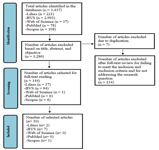
Source: Adapted from Mendes, C. et al.
Prisma Flowchart (2020)
RESULTS
A
total of 10 studies were included, of which 2 were identified in LILACS (20%),
7 in BVS (70%), and 1 in Scopus (10%). All studies found were published in
English. Regarding study type, 2 were multicenter observational studies (20%),
3 were prospective cohort studies (30%), 2 were retrospective (20%), 2 were
multicenter retrospective observational studies (20%), and 1 was a
cross-sectional study (10%). Regarding levels of evidence, 3 studies were
classified as level 3 (30%) and 7 studies were classified as level 4 (70%),
based on the categorization by the U.S. Agency for Healthcare Research and
Quality (AHRQ).
Table
1 presents the characteristics of 6 studies, including authors, year, country,
journal, study type, study objective, sample, and level of evidence. It also
identifies the results found in the studies, the specific cardiovascular
complication reported, and its incidence, in relation to patients without a
history of cardiovascular disease (CVD). Table 2 presents the same
characteristics, results, and incidence of the cardiovascular complications
listed in Table 1, but in relation to patients with a history of CVD.
The
first two studies in Table 2 are already described in Table 1 but are also
included in Table 2 because they addressed cardiovascular complications
triggered by the infection in both groups of patients with and without a
history of CVD. Table 3, on the other hand, presents the pathophysiology of
cardiovascular complications and COVID-19, which are addressed in the results,
discussion, and conclusion sections of the 8 studies that also address this
pathophysiological topic among the 10 analyzed in the study as a whole.
A
study was conducted on the prevalence and a meta-analysis of cardiovascular
complications in adults with and without a history of CVD, as reported in the
analyzed studies. Figure 1 presents the meta-analysis of the prevalence of
cardiovascular complications following SARS-CoV-2 infection in adults without a
history of CVD, based on 6 studies totaling 1,068 participants. The combined
results, using a random-effects model, indicate an estimated prevalence of
cardiovascular complications of 33%, with a 95% CI of 15% to 58% (the 95% CI
provides a range of values within which the prevalence would be expected to
fall if a larger sample size were obtained in each study), suggesting
considerable heterogeneity among the studies (I² = 98%, p < 0.01), with
individual estimates ranging from 8% to 61%
Figure
2 presents the meta-analysis of the prevalence of cardiovascular complications
following SARS-CoV-2 infection in adults with a history of CVD, based on 6
studies involving a total of 53,810 participants. The estimated pooled
prevalence, using a random-effects model, was 52%, with a 95% CI of 34% to 70%,
indicating a high prevalence of cardiovascular complications and a wide
confidence interval. Furthermore, the analysis revealed extremely high
heterogeneity among the studies (I² = 100%, p = 0), suggesting a wide variation
in the reported results. Individual prevalence estimates varied significantly,
ranging from 18% to 73%, which may be attributed to differences in the study
populations, clinical contexts, and methodologies used in the studies.
This
high heterogeneity reflects significant variations in the reported prevalences
and can be attributed to methodological or population-based differences among
the analyzed studies. The high heterogeneity observed underscores the need for
caution in interpreting the results and suggests that future studies should
explore the causes of these variations to improve our understanding of the
factors influencing cardiovascular complications in patients with COVID-19.
Figure
1
- Meta-analysis of the prevalence of cardiovascular complications following
SARS-CoV-2 infection in adults without prior cardiovascular disease. Systematic
review, Brazil, 2024.
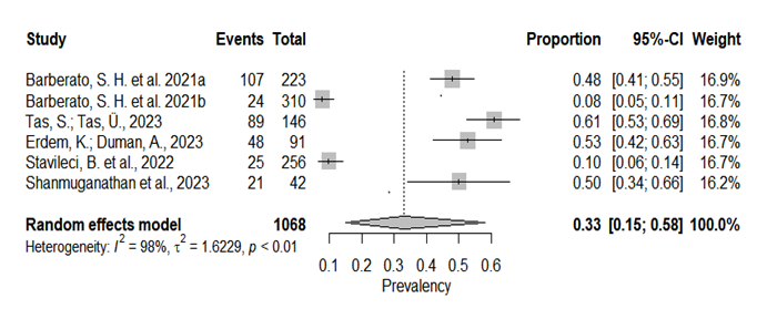
Source:
Prepared by the authors.
Figure
2
- Meta-analysis on the prevalence of cardiovascular complications following
SARS-CoV-2 infection in adults with pre-existing cardiovascular disease.
Systematic review, Brazil, 2024.
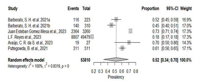
Source:
Prepared by the authors.
DISCUSSION
The SARS-CoV-2 virus
infects host cells, causing COVID-19, when the viral membrane S protein binds
to Angiotensin-Converting Enzyme 2 (ACE2) and is cleaved by Transmembrane
Serine Protease 2 (TMPRSS2), which allows the viral genome to enter the cell
and replicate; thus, for the cell to become infected, expression of the ACE2
receptor on the cell membrane is necessary, as well as TMPRSS2, ADAM17, and
CTSL—two other auxiliary proteases expressed in the cardiovascular system 21,21.
The
ECA2 receptor is present in nearly all vascular cells, endothelial cells,
fibroblasts, and pericytes, and ECA2 itself plays an active role in the
renin-angiotensin-aldosterone system, which is essential for homeostasis, blood
pressure regulation, and immune system function. Thus, at the cellular and
tissue levels, SARS-CoV-2 infection leads to a cytokine storm, cellular
oxidative stress, and consequent inflammation and endothelial dysfunction 21,21.
Among
the selected articles, there is a consensus that SARS-CoV-2 infection and
COVID-19 lead to cardiovascular manifestations and/or complications, including:
heart rate irregularities, r sinus tachycardia, reduced heart rate variability,
palpitations, chest pain, myocardial injury, enlarged right ventricular (RV)
dimensions, elevated diastolic and systolic pulmonary artery pressures,
myocardial fibrosis, right bundle branch block, atrial fibrillation, left
bundle branch block, right ventricular dysfunction, inferior vena cava
dilatation, pericardial effusion, cardiac arrhythmia, heart failure,
endothelial dysfunction or vascular wall abnormalities, acute myocardial
infarction, atrial-ventricular fibrillation, mild left ventricular (LV) systolic
dysfunction, and acute myocardial edema.
The
clinical manifestations that were most common in the analyzed studies, and
which trigger other cardiovascular complications, to be discussed in greater
detail here, are: myocardial injury, endothelial dysfunction, acute myocardial
infarction (AMI), stroke, heart failure, right ventricular (RV) dysfunction,
and left ventricular (LV) systolic dysfunction.
Myocardial
injury is characterized by elevated troponin levels above the 99th percentile,
and is usually accompanied by abnormalities on the electrocardiogram and
echocardiogram, such as wall abnormalities and left ventricular diastolic
dysfunction, right ventricular dysfunction, and the development of scarring
resembling myocarditis, and occurs in 20% to 30% of patients with COVID-1 22,23.
Myocardial injury and the resulting increase in troponin also serve as
predictors of mortality. When comparing patients with and without myocardial
injury, those who develop this complication exhibit more cardiovascular
abnormalities than patients without myocardial injury, such as changes in the
electrocardiogram and echocardiogram, hyperinflammation, and anatomical and
functional abnormalities of the ventricles 22.
Myocardial
injury generally occurs during the acute phase of the disease, either due to
direct viral damage to the myocardium via entry through the ACE2 receptor, or
due to the immune response and resulting cytokine storm, or through an indirect
mechanism resulting from respiratory failure and the induction of hypoxia in
myocardial tissue 24. Myocardial injury was observed in 60.8% of
cases, and non-ischemic myocardial injury in the form of possible myocarditis
was observed in 10.1% of cases19. fQRS is an electrocardiographic
sign of the presence of myocardial scar tissue in response to myocardial
injury; it indicates delayed ventricular conduction and the development of
heart failure and arrhythmias, and was present in 36.9% of cases 14,17.
Vascular injury, hyperinflation, myocardial injury, and necrosis alter the
heartbeat and lead to autonomic dysfunction and sympathetic hyperactivity or
parasympathetic hypoactivity, as observed by heart rate variability. Changes in
cardiac autonomy and in the sympathetic and parasympathetic activities of the
nervous system lead to a condition of atrioventricular fibrillation12.
Endothelial
dysfunction, in turn, can be explained largely by the direct consequences of
viral invasion and by the immune response to the infection. Changes in nitric
oxide availability trigger immune reactions that disrupt vascular homeostasis,
causing damage to the tunica media and an increase in the expression of prothrombotic
factors, thereby exacerbating the body’s pro-inflammatory state. Direct
infection of the endothelial cell itself causes its dysfunction and accelerates
the state of coagulopathy and thrombosis, creating a risk of stroke and
ischemic heart attacks 18,21. Endothelial dysfunction is prevalent
in 51.8% of cases, and patients with this complication have a higher incidence
of chronic heart failure (71.4%) and hypertension (78.6%) compared to patients
with preserved endothelial function19, 18.
A
relationship can be observed between endothelial dysfunction and other
cardiovascular complications identified in the studies: acute myocardial
infarction (AMI) and stroke. AMI is the ischemia and death of myocardial tissue
that can occur due to obstruction of the coronary arteries (type 1 AMI) or due
to an imbalance between oxygen demand and supply, while stroke is a
neurological deficit caused by a vascular event, such as a cerebral infarction,
and occurs in a small number of cases, approximately 0.6% 17,23.
Endothelial dysfunction and systemic inflammation lead to thrombus formation
and the clinical presentation of type 1 AMI, and this same inflammatory state
increases the myocardium’s oxygen demand, which, exacerbated by respiratory
problems and secondary hypoxia, can lead to a type 2 AMI 25.
Furthermore, ST-segment elevation myocardial infarction (STEMI) was observed in
31.5% of cases, 12.1% more prevalent than non-ST-segment elevation myocardial
infarction (19.4% of cases) 19.
Heart
failure (HF), in turn, is a syndrome resulting from cardiac abnormalities,
whether functional or structural in nature 17. It generally occurs
as a consequence of acute myocarditis or as a response to the cardiac muscle’s
oxygen demand and hyperinflammation. Dysfunction of ACE2 and elevated
angiotensin II levels are also factors that explain its pathophysiology, occurring
in 44.4% of cases among patients with pre-existing CVD 23,19. Heart
failure is a predictor of patient mortality, which is 1.6 times higher compared
to those without heart failure, and SARS-CoV-2 infection may be a risk factor
for its development at other stages of life 19, 12.
Left
ventricular (LV) systolic dysfunction is defined as a reduction of less than
50% in the left ventricular ejection fraction (LVEF) and can range from mild
(with an LVEF between 40 and 50%), moderate (between 30 and 39%), to severe
(with an LVEF less than 30%) 11. Left ventricular diastolic function
is also assessed using parameters other than LVEF, and the most accurate measure
of left ventricular diastolic function is the E/Em ratio, which is the ratio of
the peak early mitral inflow velocity (E) to the early diastolic mitral annular
velocity (Em) 12.
Changes in right ventricular (RV) dimensions are assessed by the
Myocardial Performance Index (MPI), a representative marker of overall function
that has prognostic value for many cardiac conditions. Right ventricular (RV)
structural and functional abnormalities are associated with increased
sympathetic tone and decreased HRV (heart rate variability). One hypothesis
for these abnormalities and cardiac structural changes may be pneumonia 13.
The most common manifestation in the acute phase of COVID-19 is right
ventricular dilation/dysfunction, and these changes are used as an independent
predictor of 60-day mortality and a high-risk factor for an unfavorable outcome
12,13.
There
are some differences between the cardiovascular complications that occur in
patients with and without a history of prior CVD. Left ventricular (LV)
systolic and diastolic dysfunction, as well as LV dilation and hypertrophy, are
reported in 4%, 39.4%, 7.6%, and 27.2% of cases, respectively, in patients
without a history of CVD 10. Complications such as right ventricular
(RV) systolic dysfunction are more common in patients without a history of CVD
compared to those with a history of CVD and are the most frequent complication;
and only 52% of patients without a history of CVD had a normal echocardiogram;
LV hypertrophy and pulmonary hypertension are more common in patients without
CVD, followed by right ventricular systolic dysfunction and left ventricular
systolic dysfunction 10.
It
is worthwhile to analyze studies that highlight the persistence or emergence of
new cardiovascular complications even after the acute phase of COVID-19 and in
patients with no history of cardiovascular disease (CVD). Cardiac involvement
was observed in 78% of patients who recovered from COVID-19, and the
persistence of at least one symptom months after the acute phase, with the most
commonly reported being palpitations (60.8%), chest pain (43.1%), fatigue
(47.1%), and dyspnea (27.5%). The mean systolic blood pressure and mean
diastolic blood pressure in post-COVID patients are 128.9 and 79.3,
respectively—values very similar to those observed in patients during the acute
phase, which were 130.4 and 80.5, respectively. The rate of abnormal initial
heart rate variability is higher in post-COVID-19 patients, and endothelial
function remains impaired three months after SARS-CoV-2 infection 12,18.
HRV and HRT abnormalities persisted in the post-COVID group, indicating the
persistence of cardiac autonomic dysfunction after clinical resolution;
therefore, medical follow-up is necessary even after clinical resolution 12.
Thus,
it can be observed that COVID-19 can lead to long-term cardiovascular
complications even in patients with no history of CVD and even months after the
acute infection, potentially serving as a risk factor for the development of
cardiovascular diseases, such as heart failure and hypertension 18,12.
CONCLUSION
This
study shows that SARS-CoV-2 infection affects the cardiovascular system both
directly and indirectly, with pathophysiological manifestations at the
cellular, tissue, and systemic levels. Myocardial injury, endothelial
dysfunction, and disturbances in oxygen supply and demand affect the myocardium
and exacerbate other immunological and pathological reactions in the cardiac,
respiratory, and immune systems, among others. Heart failure, right ventricular
dysfunction, systolic dysfunction of the left and right ventricles, atrial
fibrillation, and stroke are some of the most common cardiovascular
complications triggered by SARS-CoV-2 infection.
The
absence of a prior history of cardiovascular disease (CVD) does not guarantee
that SARS-CoV-2 infection will not lead to cardiovascular complications, and
there is no significant difference compared to patients with a prior history of
CVD. SARS-CoV-2 infection may even lead to future complications in patients
without a history of CVD or exacerbate pre-existing cardiomyopathies.
REFERENCES
1 Ministério da Saúde (BR).
Secretaria de Vigilância em Saúde. Guia de vigilância epidemiológica:
emergência de saúde pública de importância nacional pela doença pelo
coronavírus 2019 – covid-19. Brasília-DF: Ministério da Saúde; 2022. Disponível
em: https://www.gov.br/saude/pt-br/assuntos/covid-19/publicacoes-tecnicas/guias-e-planos/guia-de-vigilancia-epidemiologica-covid-19/view.
2 Su S, Wong G, Shi W, Liu J, Lai
ACK, Zhou J, Liu W, Bi Y, Gao GF. Epidemiology, Genetic Recombination, and
Pathogenesis of Coronaviruses. Trends Microbiol. 2016 Jun;24(6):490-502. Doi:
10.1016/j.tim.2016.03.003. Epub 2016 Mar 21. PMID: 27012512; PMCID: PMC7125511.
3 Organização Pan-Americana da
Saúde. Histórico da pandemia de COVID-19 - OPAS/OMS. OPAS; 2024. Disponível em:
https://www.paho.org/pt/historico-da-emergencia-internacional-covid-19.
4 World Health Organization. COVID-19
epidemiological update – 17 May 2024. Disponível em: https://www.who.int/publications/m/item/covid-19-epidemiological-update-edition-167.
5 Ministério da Saúde (BR).
Coronavírus Brasil. Painel COVID. Atualizado em: 08/09/2024. Disponível em: https://covid.saude.gov.br/.
6 Sharma A, Ahmad Farouk I, Lal SK.
COVID-19: A Review on the Novel Coronavirus Disease Evolution, Transmission,
Detection, Control and Prevention. Viruses. 2021 Jan 29;13(2):202. Doi:
10.3390/v13020202. PMID: 33572857; PMCID: PMC7911532.
7 Rodrigues SS, Galli RA. Análise
fisiopatológica das manifestações clínicas respiratórias em pessoas infectadas
pelo vírus SARS-CoV-2. Rev Médica de Minas Gerais. 2022;32. Doi:
10.5935/2238-3182.2022e32201
8 Carvalho FRS. et al.
Fisiopatologia Da Covid-19: Repercussões Sistêmicas. Unesc em Revista.
2020;4(2): 170–84, 2020. Doi:10.22592/ode2022n39e312
9 Martins JDN, Sardinha DM, Silva
RR, Lima KVB, Lima LNGC. As Implicações da COVID-19 no sistema cardiovacular:
prognostico e intercorrencias. J. Health Biol Sci. 2020;8(1):1-9.
Doi: 10.12662/2317-3206jhbs.v8i1.3355.p1-9.2020
10 Barberato SH, Borsoi R, Roston F, Miranda
HLM, Patriota P, Otto ME, et al. Achados Ecocardiográficos em Pacientes com
COVID-19 com e sem Doença Cardiovascular Prévia. Arq. Bras. Cardiol. nov.
2021;117(6):1207-11. Doi: https://doi.org/10.36660/abc.20201300.
11 Barberato SH, Borsoi R, Roston F,
Miranda HLM, Patriota P, Otto ME, et al. Transthoracic Echocardiography
Findings of Hospitalized Patients with COVID-19: Results from the Brazilian
Echocardiography Registry During the COVID-19 Pandemic (ECOVID). Arq Bras
Cardiol: Imagem cardiovasc. 28 mar. 2021;34(4). Doi: 10.47593/2675-312X/20213404eabc256.
12 Taş S, Taş Ü. Effects of COVID-19
on the Autonomic Cardiovascular System: Heart Rate Variability and Turbulence
in Recovered Patients. Tex Heart Inst J. 2023. Aug 21;50(4):e227952. Doi:
10.14503/THIJ-22-7952. PMID: 37605870; PMCID: PMC10660136.
13 Erdem K, Duman A. Pulmonary artery
pressures and right ventricular dimensions of post-COVID-19 patients without
previous significant cardiovascular pathology. Heart Lung. jan. 2023; 57:75–79.
Doi: https://doi.org/10.1016/j.hrtlng.2022.08.023
14 Stavileci B, Özdemir E, Özdemir B,
Ereren E, Cengiz M. De-novo development of fragmented QRS during a six-month
follow-up period in patients with COVID-19 disease and its cardiac effects. J
Electrocardiol. 2022 May-Jun;72:44-48. Doi: 10.1016/j.jelectrocard.2022.02.012.
Epub 2022 Mar 12. PMID: 35306293; PMCID: PMC8917010.
15 Shanmuganathan M,
Kotronias RA, Burrage MK, Ng Y, Banerjee A, Xie C, Fletcher A, Manley P. et al.
Acute
changes in myocardial tissue characteristics during hospitalization in patients
with COVID-19. Frente.
Cardiovascular. 2023;
Med.10:1097974.
doi: 10.3389/fcvm.2023.1097974
16 Gomez-Mesa JE,
Galindo S, Escalante-Forero M, Rodas Y, Valencia A, Perna E, Romero A, Mendoza
I, Wyss F, Barisani JL, Speranza M, Alarco W, Flórez NA. Latin-American
Registry of Cardiovascular Disease and COVID-19: Final Results. Glob Heart.
2023 Nov 1;18(1):60. doi: 10.5334/gh.1272. PMID: 37928360; PMCID: PMC10624135.
17 Reyes LF, Garcia-Gallo E, Murthy
S, Fuentes YV, Serrano CC, Ibáñez-Prada ED, et al. ISARIC Characterisation Group.
Major adverse cardiovascular events (MACE) in patients with severe COVID-19
registered in the ISARIC WHO clinical characterization protocol: A prospective,
multinational, observational study. J Crit Care. 2023 Oct;77:154318. Doi:
10.1016/j.jcrc.2023.154318. Epub 2023 May 9. PMID: 37167775; PMCID:
PMC10167415.
18 Araújo CRDS, Fernandes J,
Caetano DS, Barros AEVDR, de Souza JAF, Machado MDGR, de Aguiar MIR, Brandão
SCS, Campos SL, de Andrade AFD, Brandão DC. Endothelial function, arterial
stiffness and heart rate variability of patients with cardiovascular diseases
hospitalized due to COVID-19. Heart Lung. 2023 Mar-Apr;58:210-216. Doi:
10.1016/j.hrtlng.2022.12.016. Epub 2023 Jan 2. PMID: 36621104; PMCID:
PMC9805900.
19 Puttegowda B, Shivashankarappa
AB, Dutta S, Chikkamuniswamy R, Bhat P, Krishnan S, Nanjappa MC. Patterns of
cardiovascular diseases in COVID-19 patients admitted to tertiary cardiac care
centre. Indian Heart J. 2021 Nov-Dec;73(6):682-686. Doi:
10.1016/j.ihj.2021.10.007. Epub 2021 Oct 22. PMID: 34695446; PMCID: PMC8532374.
20 Chen C, Wang J,
Liu YM. Single-cell analysis of adult human heart across healthy and
cardiovascular disease patients reveals the cellular landscape underlying
SARS-CoV-2 invasion of myocardial tissue through ACE2. J Transl Med. 2023 May
31;21(1):358.
Doi: https://doi.org/10.1186/s12967-023-04224-1
21Pelisek J, Reutersberg B, Greber
UF, Zimmermann A. Vascular dysfunction in COVID-19 patients: update on
SARS-CoV-2 infection of endothelial cells and the role of long non-coding RNAs.
Clinical Science. Nov. 2022;136(21):1571–90. Doi: 10.1042/CS20220235. PMID:
36367091; PMCID: PMC9652506.
22 Shu H, Wen Z, Li N, Zhang Z,
Ceesay BM, Peng Y, Zhou N, Wang DW. COVID-19 and Cardiovascular Diseases: From
Cellular Mechanisms to Clinical Manifestations. Aging Dis. 2023 Dec
1;14(6):2071-2088. Doi: 10.14336/AD.2023.0314. PMID: 37199573; PMCID:
PMC10676802.
23 Armas E, Sucet K, Mola M,
Katsuyori; Batista Cuenca, Milton. El sistema cardiovascular y el daño inducido
por la infección por coronavirus-2. Rev Cubana Medicina. 2020;60(2):
e1638.
Disponível em: https://revmedicina.sld.cu/index.php/med/article/view/1638.
24 Terzic CM, Medina-Inojosa BJ.
Cardiovascular Complications of Coronavirus Disease. Phys Med Rehabil
Clin N Am.
2023; 34(3): 551-61.
Doi:
https://doi.org/10.1016/j.pmr.2023.03.003.
25 Elias-Sierra R, Elias-Armas KS,
González-Tirado F, Maceo-Sobrado E. Influencia de la COVID-19 sobre el sistema
cardiovascular. Gac Med Est [en línea]. 2020; 1(2):133-45.
Funding
and Acknowledgments:
The
research received no funding. Conflict of Interest Statement: “None to
declare.” Data Availability Statement: No datasets were generated in this study.
The information presented is described in the body of the article.
Authorship
Criteria (author contributions):
Vitória
Pereira Silva Dutra; Solon Alves da Silva Filho; Antonio Frivaldo Marinho Neto;
Marcos Vinícius Novais Alves; Rafael Aires Oliveira Campos:
Contributed to data acquisition, analysis, and/or interpretation;
Karla
Vanessa Morais Lima; Arlane Silva Chaves; Francisco Alves Lima Júnior:
Contributed substantially to the study conception and/or design, as well as to
the drafting, critical revision, and final approval of the published version.
Scientific
Editor: Ítalo Arão Pereira Ribeiro. ORCID:
https://orcid.org/0000-0003-0778-1447
Rev Enferm Atual In Derme 2026;100(3):
e026104