REVIEW ARTICLE


ULTRASOUND-GUIDED VENOUS PUNCTURE IN URGENT AND EMERGENCY CARE: A SCOPING REVIEW


PUNÇÃO VENOSA GUIADA POR ULTRASSONOGRAFIA EM URGÊNCIA E EMERGÊNCIA: REVISÃO DE ESCOPO


PUNCIÓN VENOSA GUIADA POR ULTRASONIDO EN URGENCIAS Y EMERGENCIAS: REVISIÓN DE ALCANCE


https://doi.org/10.31011/reaid-2026-v.100-n.3-art.2790


Ana Flávia Gomes de Vargas e Lima1

Amanda Dias de Souza1,2

Juliana de Melo Vellozo Pereira Tinoco1,3

Ana Carla Dantas Cavalcanti1,4


1Programa Profissional de Enfermagem Assistencial, Universidade Federal Fluminense (UFF). Rio de Janeiro – Rio de Janeiro, BraZil. 1ORCID: https://orcid.org/0009-0005-2179-545X

2Orcid: https://orcid.org/0009-0005-4683-4558

3Orcid: https://orcid.org/0000-0002-2418-6984

4Orcid:https://orcid.org/0000-0003-3531-4694


Corresponding Author

Ana Flávia Gomes de Vargas e Lima

Rua: Dr. Celestino, 74 - Centro, Niterói - Rio de Janeiro. Brazil. CEP: 24020-094.  phone: +55(31) 98700-1459, E-mail: af_lima@id.uff.br


Submission: 28-04-2026

Approval: 08-06-2026


ABSTRACT

Introduction: Although there is consistent evidence regarding the benefits of ultrasonography in peripheral venous puncture, an integrated understanding of how the technique is used in practice remains limited. Objective: To map the available evidence on ultrasound-guided peripheral venous puncture performed by healthcare professionals in urgent and emergency care settings, considering clinical practices, training strategies, protocols, barriers, facilitators, and associated outcomes. Method: A scoping review conducted in accordance with the Joanna Briggs Institute methodology and reported following PRISMA-ScR guidelines. The search was performed in PubMed/MEDLINE, Embase, LILACS, Scopus, and Web of Science, with no language or time restrictions. Studies addressing the use of ultrasonography for peripheral venous puncture were included. Study selection was carried out by independent reviewers, and data were analyzed through narrative synthesis. Results: A total of 27 studies were included, predominantly observational in design and concentrated in the United States. The technique was mainly used in patients with difficult venous access and was associated with higher first-attempt success rates, reduced number of punctures, shorter procedure time, and fewer complications. Effectiveness was associated with professional training and institutional support. Barriers included limited equipment availability, lack of standardized protocols, and variability in training, as well as a gap in the literature regarding the use of the technique in prehospital settings. Conclusion: The consolidation of ultrasound-guided peripheral venous puncture depends on professional training, protocol standardization, and organizational support, highlighting the need for further studies across different healthcare settings.

Keywords: Ultrasonography; Catheterization, Peripheral; Venous puncture; Emergency Medical Services; Nursing.


RESUMO

Introdução: Embora existam evidências consistentes sobre os benefícios da ultrassonografia na punção venosa periférica, permanece limitada a compreensão integrada sobre como a técnica é utilizada na prática. Objetivo: Mapear as evidências disponíveis sobre a punção venosa periférica guiada por ultrassonografia realizada por profissionais de saúde em contextos de urgência e emergência, considerando práticas assistenciais, estratégias de treinamento, protocolos, barreiras, facilitadores e desfechos associados. Método: Revisão de escopo conduzida conforme a metodologia do Joanna Briggs Institute e reportada segundo o PRISMA-ScR. A busca foi realizada nas bases PubMed/MEDLINE, Embase, LILACS, Scopus e Web of Science, sem restrição de idioma ou período. Foram incluídos estudos que abordassem o uso da ultrassonografia para punção venosa periférica. A seleção foi realizada por revisores independentes e os dados foram analisados por síntese narrativa. Resultados: Foram incluídos 27 estudos, com predominância de delineamentos observacionais e concentração nos Estados Unidos. A técnica foi predominantemente utilizada em pacientes com acesso venoso difícil e esteve associada a maior taxa de sucesso na primeira tentativa, redução do número de punções, menor tempo de procedimento e menor ocorrência de complicações. A efetividade mostrou-se associada à capacitação profissional e ao suporte institucional. Foram identificadas barreiras relacionadas à disponibilidade de equipamentos, ausência de protocolos e variabilidade na formação, além de lacuna na literatura quanto ao uso da técnica no contexto pré-hospitalar. Conclusão: A consolidação da punção venosa periférica guiada por ultrassonografia depende da capacitação profissional, da padronização de protocolos e do suporte organizacional, sendo necessária a ampliação de estudos em diferentes contextos assistenciais.

Palavras-chave: Ultrassonografia; Cateterismo Periférico; Punção Venosa; Serviços Médicos de Emergência; Enfermagem.


RESUMEN

Introducción: Aunque existen evidencias consistentes sobre los beneficios de la ultrasonografía en la punción venosa periférica, persiste una comprensión integrada limitada sobre cómo se utiliza la técnica en la práctica. Objetivo: Mapear las evidencias disponibles sobre la punción venosa periférica guiada por ultrasonografía realizada por profesionales de la salud en contextos de urgencias y emergencias, considerando prácticas asistenciales, estrategias de capacitación, protocolos, barreras, facilitadores y desenlaces asociados. Método: Revisión de alcance realizada conforme a la metodología del Joanna Briggs Institute y reportada según la guía PRISMA-ScR. La búsqueda se llevó a cabo en las bases de datos PubMed/MEDLINE, Embase, LILACS, Scopus y Web of Science, sin restricción de idioma ni período. Se incluyeron estudios que abordaran el uso de la ultrasonografía para la punción venosa periférica. La selección fue realizada por revisores independientes y los datos fueron analizados mediante síntesis narrativa. Resultados: Se incluyeron 27 estudios, con predominio de diseños observacionales y concentración en los Estados Unidos. La técnica se utilizó principalmente en pacientes con acceso venoso difícil y se asoció con una mayor tasa de éxito en el primer intento, reducción del número de punciones, menor tiempo de procedimiento y menor ocurrencia de complicaciones. La efectividad se asoció con la capacitación profesional y el apoyo institucional. Se identificaron barreras relacionadas con la disponibilidad de equipos, la ausencia de protocolos estandarizados y la variabilidad en la formación, además de una laguna en la literatura respecto al uso de la técnica en el contexto prehospitalario. Conclusión: La consolidación de la punción venosa periférica guiada por ultrasonografía depende de la capacitación profesional, la estandarización de protocolos y el apoyo organizacional, siendo necesaria la ampliación de estudios en diferentes contextos asistenciales.

Palabras clave: Ultrasonografía; Cateterismo Periférico; Punción venosa; Servicios Médicos de Urgencia; Enfermería.


INTRODUCTION

Peripheral venous access is an essential procedure in urgent and emergency care settings, widely used for the timely administration of fluids, medications, and other intravenous therapies. However, establishing access can be particularly challenging in patients with difficult venous access, such as those with obesity, a history of multiple punctures, a history of intravenous drug use, or debilitating chronic conditions, leading to repeated attempts, delayed treatment initiation, and increased patient discomfort and procedure-related complications(1-6).

In this context, point-of-care ultrasound has been adopted as a strategy to assist with peripheral venous access, enabling direct visualization of vascular structures and greater precision during catheter insertion. Evidence shows that, in patients with difficult venous access, ultrasound-guided techniques are associated with higher first-attempt success rates, fewer punctures, shorter procedure times, and greater patient satisfaction compared to the conventional approach(2,3,7).

 Beyond the benefits regarding procedural effectiveness, the literature indicates that the performance of ultrasound-guided peripheral venous access depends on multiple factors, including vessel anatomy, the patient's clinical condition, the professional's experience, and service organization. Studies demonstrate that variables such as vessel visibility and trajectory directly influence puncture success, even when image guidance is used, underscoring the need to integrate technology, clinical judgment, and technical skill(6).

 Another key aspect is professional training. Adopting ultrasound for peripheral venous access requires the development of specific competencies; structured training, incorporating simulation and supervised practice, is crucial for improving performance and proficiency over time. Evidence indicates a consistent rise in success rates as operator experience grows, pointing to a learning curve associated with the technique(8,9). Despite growing recognition of its benefits, the adoption of ultrasound-guided peripheral venous access varies widely across healthcare services and professionals. The literature describes variations in equipment availability, the existence of institutional protocols, training levels, and the technique's adoption in clinical practice, factors that can limit its effectiveness and sustainability(10).

 Furthermore, scientific literature on the subject displays significant methodological heterogeneity, encompassing clinical trials, observational studies, qualitative research, and implementation studies. Although there is consistent evidence regarding the benefits of ultrasound for peripheral venous access, there remains a limited integrated understanding of how the technique is utilized in practice, the training strategies employed, the barriers and facilitators influencing its adoption, and the outcomes reported across different care settings.

This gap is particularly relevant in urgent and emergency care settings, where rapid and effective venous access is critical for clinical decision-making and the quality of care. In this context, scoping reviews are appropriate for mapping the extent, nature, and characteristics of available evidence, especially in areas marked by methodological diversity and knowledge gaps, thereby enabling the identification of patterns, inconsistencies, and research priorities(11).

 Given this, the present study aimed to map the available evidence regarding ultrasound-guided peripheral venous cannulation performed by healthcare professionals in urgent and emergency settings, considering clinical practices, training strategies, protocols, barriers, facilitators, and associated outcomes.


METHODS

This is a scoping review conducted according to the methodology proposed by the Joanna Briggs Institute (JBI) and reported in accordance with the Preferred Reporting Items for Systematic Reviews and Meta-Analyses extension for Scoping Reviews (PRISMA-ScR) checklist (11,12). The review protocol was previously registered on the Open Science Framework (OSF) under DOI 10.17605/OSF.IO/VP6GT, outlining the objectives, research question, eligibility criteria, search strategies, source selection procedures, data extraction process, and synthesis method, thereby ensuring transparency and methodological reproducibility.

 The research question was structured based on the PCC (Population, Concept, and Context) framework, which is recommended for scoping reviews (13). The population was defined as healthcare professionals; the concept as ultrasound-guided peripheral venous access, encompassing clinical practices, training strategies, protocols, barriers, facilitators, and associated outcomes; and the context as urgent care, hospital emergency services, and pre-hospital care settings. Based on this structure, the following guiding question was defined: how does the scientific literature describe and characterize the use of ultrasound-guided peripheral venous access by healthcare professionals in these care settings?

 Sources of evidence included publications addressing the use of ultrasound for peripheral venous access performed by healthcare professionals in the defined settings, including studies describing clinical practices, training strategies, protocols, clinical experiences, barriers, facilitators, or outcomes related to the technique. Consistent with the nature of scoping reviews, there were no restrictions regarding methodological design; eligible sources included quantitative, qualitative, and mixed-methods primary studies, as well as implementation studies and experience reports relevant to mapping the topic. There were no restrictions regarding language or publication date. Studies focusing on topics unrelated to the defined concept and context were excluded, as were publications dedicated exclusively to diagnostic ultrasound, studies on vascular access types unrelated to the review's objective, editorials, letters to the editor, commentaries, and abstracts lacking full-text availability. Systematic reviews and scoping reviews were not included as primary sources of evidence but were used solely for manual reference screening.

 The search strategy was developed in a structured and iterative manner, utilizing controlled descriptors from the Medical Subject Headings (MeSH) and Health Sciences Descriptors (DeCS) vocabularies, combined with free-text terms, synonyms, Boolean operators (AND, OR), and truncation symbols, tailored to the specific requirements of each database. Strategy development followed sequential steps: identifying key terms related to the PCC framework components, converting these terms into controlled vocabulary, combining descriptors with free-text keywords, and adapting the syntax for each indexing database. Searches were conducted in PubMed/MEDLINE, Embase, LILACS, Scopus, and Web of Science, databases selected for their relevance to the fields of health and nursing. The initial search was performed in May 2025 and updated on July 11, 2025, to incorporate more recent studies. The complete search strategies used for each database, along with the number of records retrieved, are detailed in Chart 1.


Chart 1 – Search strategies for the scoping review based on the PCC strategy

Estage

P – Population

C – Concept

C – Context

Extraction

Healthcare professionals

Practices related to

ultrasound-guided peripheral venipuncture

Urgent and emergency services,


mobile pre-hospital care units


Conversion and combination

MeSH Terms: Health Personnel

Outros termos: Health professional; nurse*; paramedic*

physician*

MeSH Terms:

Catheterization; Peripheral


Outros termos: Catheter*; cannulat*; intraven*; venipuncture; ultrasound*;

POCUS

MeSH Terms: Emergency Medical Services; Emergency Service, Hospital; Ambulances

Outros termos: Emergenc*; urgenc*; prehosp*

Syntax strategies using Boolean operators

Databases/


repository

Articles found

("Health Personnel"[MeSH Terms] OR health professional OR nurse* OR paramedic* OR physician*) AND (("Catheterization, Peripheral"[MeSH Terms] OR catheter* OR cannulat* OR intraven* OR venipuncture) AND (ultrasound* OR POCUS)) AND ("Emergency Medical Services"[MeSH Terms] OR "Emergency Service, Hospital"[MeSH Terms] OR "Ambulances"[MeSH Terms] OR emergenc* OR urgenc* OR prehosp*)

PubMed/

MEDLINE

443

('health care personnel'/exp OR nurse* OR paramedic* OR physician* OR health professional*) AND (('peripheral venous catheter'/exp OR catheter* OR cannulat* OR intraven* OR venipuncture*) AND ('ultrasonography'/exp OR ultrasound* OR pocus)) AND ('emergency health service'/exp OR 'emergency medical service'/exp OR 'ambulance'/exp OR emergenc* OR urgenc* OR prehosp*)

EMBASE

195

("Personal de Salud" OR “profesional de la salud” OR enfermer* OR paramédic* OR médic*) AND (("Cateterismo Periférico" OR catéter* OR cánul* OR intraven* OR venopunción*) AND ("Ultrasonografía" OR ultrasonido* OR POCUS OR "ultrasonido en el punto de atención")) AND ("Servicios Médicos de Urgencia" OR “ambulâncias” OR servicio de urgencia OR emergenc* OR urgenc* OR prehospitalar)

LILACS

607

TITLE-ABS-KEY health AND professional* OR nurse* OR paramedic* OR physician* OR "health personnel" ) AND ((TITLE-ABS-KEYcatheter* OR cannulat* OR intraven* OR venipuncture*) AND TITLE-ABS-KEY (ultrasound* OR pocus))) AND TITLE-ABS-KEY (emergenc* OR urgenc* OR prehosp* OR ambulance* OR "emergency service" OR "emergency medical service")

Scopus

150

TS=(health professional* OR nurse* OR paramedic* OR physician* OR "health personnel") AND ( TS=(catheter* OR cannulat* OR intraven* OR venipuncture*) AND TS=(ultrasound* OR POCUS OR "point-of-care ultrasound")) AND TS=(emergenc* OR urgenc* OR prehosp* OR ambulance* OR "emergency service" OR "emergency medical service")

Web of Science

400


Additionally, a manual search of the reference lists of the included studies was conducted to identify potentially relevant publications not retrieved during the electronic search. No systematic search of gray literature was performed; this may limit the scope of the mapping and should be considered when interpreting the results.

 The retrieved records were exported to Rayyan® software, where duplicates were identified and removed(11). The process of selecting sources of evidence took place in two sequential stages. In the first stage, titles and abstracts were screened against previously defined eligibility criteria. In the second stage, potentially relevant studies underwent full-text review to verify final eligibility. Both stages were conducted by two independent reviewers who had previously aligned on inclusion and exclusion criteria. Disagreements were resolved by consensus and, when necessary, by a third reviewer. Reasons for exclusion during the full-text review stage were systematically recorded. The study selection flow was organized according to the PRISMA-ScR diagram, ensuring the process was transparent and auditable.

 Data extraction was performed using a standardized form that had been previously developed and pilot-tested on selected studies, in accordance with JBI methodological recommendations(11). The form covered information regarding general study characteristics, such as authorship, publication year, and country of origin, as well as methodological design, the population and professional categories involved, the care setting, practices related to ultrasound-guided peripheral venous cannulation, reported clinical and operational outcomes, and barriers and facilitators associated with the technique's adoption. The extraction process was carried out by independent reviewers with cross-checking of data; discrepancies were resolved by consensus.

 Data synthesis was conducted using a narrative approach, organizing findings into thematic categories that included study characteristics, care practices, procedure-related outcomes, and factors influencing implementation. This approach enabled the mapping of available evidence, aligning with the objective of scoping reviews to describe the extent, nature, and characteristics of existing literature. No assessment of the methodological quality of the included studies was conducted, as is standard practice for this type of review, since the aim was not to evaluate effects or establish a hierarchy of evidence, but rather to identify and organize available knowledge on the topic.


RESULTS

Evidence source selection process


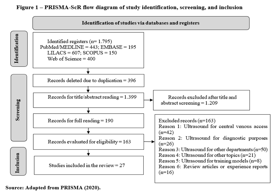
A total of 1,795 records were identified in the databases. After removing 396 duplicates, 1,399 records underwent title and abstract screening, resulting in the exclusion of 1,209 studies. Of the 190 records assessed in full text, 163 were excluded for failing to meet eligibility criteria, resulting in a total of 27 studies included in the review. The process of identifying, screening, determining eligibility for, and including evidence sources is presented in the PRISMA-ScR flow diagram (Figure 1).
Characteristics of the included studies


The included studies were published between 2004 and 2025, with a concentration in the last ten years (66.7%; n=18). Regarding geographical distribution, there was a predominance of studies conducted in the United States (74%; n=20), followed by Brazil (n=3), Australia (n=3), and Turkey (n=1). Concerning methodological design, observational studies predominated (74%; n=20), followed by randomized clinical trials (19%; n=5) and multicenter studies (7%; n=2). Detailed study characteristics, including the care setting, professionals involved, design, strategies adopted, and reported outcomes, are summarized in Chart 2.



Chart 2 - Key characteristics of the selected articles

Landscape/

Professionals

Author/

Year /

Country

Study designs/


Population

Practices/strategies

Outcome

Level 1 Trauma Center


Nurses


Brannam et al., 2004(2)

EUA

EOP

Nº: 321

Training

Assertiveness: 280 (87%)

ED


Physician

Costantino et al., 2005(3)

EUA


EO

Nº: 60

Real-time training

Assertiveness:

GI: 97%

GC: 33%


Punction time:

GI: 13 minutes

GC: 30 minutes

 Pacient satisfaction:

GI: 8,7

GC: 5,7

ED


nurses


Chinnock et al., 2007(14)

EUA

EOP

Nº 100

 Training

Assertiveness: 63%

Basilic vein: 39/55 (71%)

Brachial vein: 19/46 (41%).

ED


Nurse and paramedics

Resnick et al., 2008(15)

EUA

ECR

N:101

 LAMP puncture technique

First-time accuracy:

81,3%


Punction time:

LAMP: 4-10,5(average of 4 min)

LAP  1,6-7 (average of 2,9 min)

p = 0,004).

ED


Physicians

Bair et al., 2008(16)

EUA

ECR

Nº:44

Training


First-time accuracy:

GI:  57% (13/23)

GC: 57% (12/21)

ED


Technicians

Bauman et al., 2009(4)

EUA

EOP

Nº: 75

Institutional support

Ultrasound available

First-time accuracy:

GI: 33/41 (80,5%),

GC: 24/34 (70,6%)

Punction time:

GI: 1,3

GC: 3,1

Medical Intervention:

GI: 7,3%

GC: 20,6%

Skin punctions:

GI: 1,6

GC: 3,6


Complications:

GI: 41,5%

GC: 64,7%


Pacient satisfaction:

GI: 7,7

GC: 4,4

p = 0,0001

DEP

Trained professionals

Oakley et al., 2010(17)

Austrália

EOP

Nº: 84

Application in a real-world clinical setting

Assertiviness:

GI:  42%

GV: 38%

P=0,08


Dificult vein access:

GI: 35%

GC: 18%

P=0,003

Time of punction:

GI:2,15min

GC:4min

p < 0,001

ED

Physicians

Costantino et al., 2010(18) EUA

ECR

Nº: 60

Ultrasound available


Trained operators

Assertiviness:

GI: 89%

GC: 93%

p=0,88

ED

Nurses and Physicians

Weiner et al., 2013(19)

EUA

EMP

Nº = 50

GI= 29

GC= 21

Training

Medical assistance:

GI:

11/21(52,4%)

GC: 07/29 (24,1%)

P=0,04

Time of punction:

GI: 27,6min

GC:

26,4min

p=0,88

Number of punction:

GI: 2,0

GC: 2,1 p=0,70


Patient satisfation:

GI:86,2

GC:63,2

p=0,06

ED

Physicians,

Nurses and

Paramedics

Oliveira et al., 2016(20)

EUA

EOP

Nº:71

Training


Similar success among professionals (p = 0,13)

UPA

Nurses

Oliveira et al.,

2015(21)

BRA

ET

Nº: 200

Training


Assertiviness:

GI:

Visible veins:43% (p=0,02752)

veins with rectilinear appearance: 2.28 times larger (p=0,006);


GC:


fragile vein: 43% lower (p=0.002094),

mobile vein: 41% lower (p=0.02659),

veins with a straight course: 33% higher (p=0.04924),

palpable veins: 1.99 times higher (p=0.01329)

ED

Nurses

İsmailoğlu et al., 2014(5)

Turquia

ECR

Nº: 60

Training

Assertiviness:

GI: 70%

GC: 30%

p < 0,05

Pain:

GI:  4,77 ± 1,74

GC: 6,00 ± 1,98

ED

Nurses

Bahl, et al., 2016(7)

EUA

ECR

Nº: 124

Training


Ultrasound available

Accuracy on the first attempt:

GI: 76%

GC: 56%

p = 0,02

Time of punction:

GI: 15,8

GC: 20,7

P=0,75

ED

Nurses and Paramedics

Stolz et al., 2016(22)

EUA

EOP

Nº: 796

Training

Assertiviness 88,24%

ED

Techinicians


Duran-Gehring et al., 2016(23)

EUA

EOR

Nº: 830

Training

Accuracy:


First attempt: 86.8%


Second attempt: 11,6%

Third: 1,6%

Technicians: 0.970


Nurses: 0.97


Doctors: 0.87

DEP Nurses

Ng et al., 2017(24)

EUA

ET

Nº: 18

Training


Ultrasound available


Acceptance of the self-guided technique by two people: 65%

UPA

Nurses

Oliveira et al.,

2017(6)

BRA

ETA

Nº: 97

Training Institutional support

Ultrasound available

First-attempt success rate:


Visible veins: 43%


Straight veins: 128%

ED

Médicos e Enfermeiros

Davis et al.,

2021(25)

EUA


EOR

Nº: 147.260

Training

do not apply

ED

Nurses

Feinsmith et al., 2018(9)

EUA

EOP

Nº: 34

Training


Supervision

Accuracy:


Attempts 1 to 10 (81%)


Attempts 21 to 30 (96%)


Number of attempts:


2% reduction, P = 0.013.


DIVA: 7% reduction, P = 0.003.

ED

Nurses and Paramedics

Acuña et al., 2020(26)

EUA

EOR

Nº: 483

Ultrasound available


Trained operators

Success rate


Success: 92% (95% confidence interval [CI], 89%–94%);


First attempt: 84% (95% CI, 80%–87%)


Without complications: 396 of 483 [82%]

ICU and UPA

Enfermeiros

Oliveira et al., 2016(8)

BRA

ETD

Nº: 14

UTI:8

UPA:6

Training


Motivation

Assertiviness:

UTI: 98,5%

UPA: 100%

DEP

Nurses

Blick et al.,

2021(27)

EUA

EOR

Nº:83

Training


Supervision


First-attempt success rate: 87% (3513/4053)


Without training: 67%


With training: 83%


Complications: 25% (904/3646)

DEP

Nurses

Anderson et al., 2022(28)

EUA

EOP

Nº:35

Institutional support


Training

Accuracy: 70%.


Adherence: 40%

DEP

Medical residents


Consultants


Nurses

Lam, 2023(29)

Austrália

ET

No.: 80


Consultants: 22


Residents:25


Emergency residents: 31


Nurses:2

Emphasis on POCUS


Use of the DIVA scale

Assertiviness:

GC: 40–50%


POCUS use:


9 physicians (86.4%),


28 resident physicians (90.3%)


16 resident physicians (64.0%)


67–80% used POCUS in <25% of cases

DEP

Nurses

Jamal et al., 2023(30)

Austrália

EOP

Nº:12

Training


Trained Operators


Ultrasound available

Accuracy: 193 out of 210 (91.9%)


First attempt: 86.5%


First two attempts: 98.96%


Level I trauma center


Nurses

McKinley et al., 2024(31)

EUA

EOP

Nº: 16

Simulation-based training


Supervision

Assertiviness:

80% (160/200)


ED

Paramedics

Wasiak et al., 2025(10)

EUA

ERM

Nº:1368

Training


Institutional support


Valuing POCUS

Accuracy: 95.72% (1297)


1st attempt: 96.38% (1250)


2nd attempts: 3.32% (43)


3rd attempts: 0.31% (4)

Complications: 24%

Approach:


Out-of-plan: 83.77% (1,146)


In-plan: 16.23% (222)

Insertion site:


Forearm: 57.7% (749)


Outcomes related to ultrasound-guided peripheral venous access


A cross-sectional analysis of the studies revealed a predominance of outcomes related to the procedure's effectiveness. Key findings included an increased first-attempt success rate (37%; n=10), reduced procedure time (18%; n=5), and improved patient satisfaction linked to reduced pain (15%; n=4), particularly in settings where structured ultrasound-guided peripheral venous access programs had been implemented. Additionally, the majority of studies (93%; n=25) demonstrated greater professional proficiency when the technique was paired with structured training involving simulation and supervised practice.


Barriers to the implementation of ultrasound-guided peripheral venous access


Barriers related to technical, organizational, and training aspects were identified. Technical factors included accidental arterial punctures (3,22), initial difficulties among professionals in performing the technique (2,22,23), and limitations regarding adequate vein visualization via ultrasound (14,15). Organizational issues included a lack of institutional protocols (19,24), resource constraints (20,26), limited availability of appropriate equipment (25,29), and institutional resistance to adopting the technology (8,24). Training-related challenges included insufficient training (2,23,24), low practice adherence (6,9), and the absence of structured clinical escalation processes (19,25). Furthermore, methodological limitations were identified in the included studies—such as small sample sizes (2,16,17) and a lack of detailed analysis regarding attempts and failures, which limits the generalizability of the findings. Individual variability in skill acquisition was also observed, with only a portion of participants meeting proficiency criteria, reinforcing the need for ongoing training(22,23,27,28).


Implications for clinical practice


The studies analyzed demonstrated that ultrasound-guided peripheral venous cannulation is associated with high success rates(2-7,8,9,15-19,20-23,25-28,30-32), a low incidence of complications(4,10,26,27), a higher likelihood of first-attempt success(4,6,7,10,15-17,23,26,27,30), reduced procedure time(3,4,17,19), and fewer required punctures(8,9,16,17,19,20,24,25,26,29), all of which contribute to reduced patient discomfort(5) and the optimization of care resources. The evidence highlighted the pivotal role of ongoing training(2-9,10,16,18-25,27-30-32), particularly when combined with simulation(31), in the professional development of various healthcare roles(18,26,30), including nurses and paramedics. The implementation of structured programs was associated with increased acceptance of the technique(10,20,23,24,28,29,32), greater professional confidence, and improved clinical performance. Furthermore, the use of ultrasound contributed to reducing the need for invasive procedures(4,19), expanding the scope of multidisciplinary practice, and reinforcing the need for institutional protocols(4,6,10,28). However, studies highlighted the need for continuous monitoring of results(9,22,23,28,30,31) and for consistent professional training strategies(2,4,5,7-9,19,20,22,23,25,27,30-32) to ensure the sustainability of the practice.


Synthesis of findings


The analysis of the evidence sources allowed for a comprehensive mapping of the characteristics of ultrasound-guided peripheral venous access in urgent and emergency care settings, thereby meeting the proposed objective. The results demonstrated the use of the technique in populations with difficult venous access, the adoption of heterogeneous care practices, consistently favorable outcomes, and the influence of technical, organizational, and training-related factors on its implementation. Furthermore, significant gaps in the literature were identified, particularly regarding the limited representation of the pre-hospital setting and the lack of standardization in implementation strategies, indicating a need for further scientific research on the subject.


DISCUSSION

The integration of ultrasound into peripheral venous cannulation represents a significant advancement in the quality of care within urgent and emergency settings, with implications that extend beyond immediate clinical outcomes to encompass organizational, educational, and patient safety dimensions(4,22,8,20,27,31). The mapping reveals that, although the technique's benefits are consistently documented in the literature, research output remains concentrated in specific international contexts with limited representation from Brazil, thereby restricting the understanding of its applicability across diverse care settings.

 Findings consistently demonstrate the superiority of ultrasound-guided peripheral venous cannulation for patients with difficult venous access, showing increased success rates, fewer attempts, and reduced procedure times(2,3,5,8-10,14,17,18,21-23,26,28,29-32). Furthermore, the reduction in complications and improved patient experience reinforce the technique's potential as a strategy aligned with patient safety principles and evidence-based practice(3,4,19,10,26,27).

 However, the predominance of observational studies and the smaller proportion of randomized clinical trials(2-4,9,14,17,20,22,23,25-28,30,31) indicate that the robustness of the evidence is still limited by potential biases, including confounding factors and operator variability. Heterogeneity in study designs, outcome measures, and training processes hampers comparability across studies and limits the generalizability of results, placing some of the evidence at intermediate levels within the scientific hierarchy(33,34). In this context, there is a need for more studies characterized by greater methodological rigor and designs that incorporate contextual and organizational variables. The adoption of the technique occurs gradually and unevenly, demonstrating that its incorporation into clinical practice does not depend solely on technological availability. Structural barriers, such as equipment limitations and the absence of institutional protocols, combined with organizational and cultural factors—such as resistance to change and variability in professional adherence, constitute critical elements affecting its implementation(2,4,8,19,20,23,25,27,30,31). These findings indicate that ultrasound should be understood not merely as a technical tool but as a complex intervention, the effectiveness of which depends on the integration of technology, work processes, and institutional governance.

 In the clinical care setting, the use of ultrasound as a second-line strategy, resorted to only after multiple failed attempts with conventional methods(2,7,15,21,25,29), reveals an underutilization of the technology and limits its potential impact. Delayed adoption increases procedure time, patient discomfort, and treatment delays, suggesting a need to revise clinical protocols to incorporate the technique at an earlier stage, particularly for high-risk populations.

 Professional training emerges as a cornerstone for consolidating this practice. Evidence regarding the learning curve, along with the link between structured training, simulation, and supervised practice and improved outcomes(9,22,23,27,30,31), indicates that technical competence is built progressively and relies on systematized educational strategies. In this regard, institutionalizing continuous training programs—featuring performance assessment and competency certification, is essential to reducing variability and ensuring the quality of care.

 The limited incorporation of the technique in the pre-hospital setting represents one of the primary gaps identified. The scarcity of studies in this context(10) suggests additional challenges related to logistics, equipment portability, training, and the operational dynamics of mobile medical services. This gap highlights the need for specific investigations that take into account the particularities of this scenario and broaden the understanding of the applicability of ultrasound in out-of-hospital settings.

The implications of the findings indicate that the consolidation of ultrasound-guided peripheral venous access requires an integrated approach involving professional training, protocol standardization, and institutional support(15,22,23,26,27). Coordinating these elements can facilitate the transition from sporadic technology use to an established standard of care, aligned with patient safety and evidence-based practice.

The limitations of this review include the methodological heterogeneity of the studies, which restricts direct comparisons; the absence of a search for gray literature, which may have limited the scope of the evidence; and the predominance of studies conducted in hospital settings, which limits generalizability to other care contexts. The failure to assess methodological quality, while consistent with the objective of a scoping review, precludes inferences regarding the strength of the evidence.


CONCLUSIONS

Ultrasound-guided peripheral venous access represents a care technology with significant potential to enhance the quality of care in urgent and emergency settings, positively impacting procedure effectiveness, patient safety, and healthcare service efficiency. Findings indicate that its use is associated with better clinical and operational outcomes, particularly in patients with difficult venous access.

 However, its consolidation into clinical practice depends on overcoming structural, organizational, and educational barriers, including expanding access to equipment, implementing institutional protocols, and developing structured professional training programs. Integrating these elements is essential to ensure the sustainable adoption of the technique across various care settings.

The analysis reveals significant gaps—particularly regarding the pre-hospital context and the standardization of implementation strategies—highlighting the need for further research to broaden understanding of the technique's applicability in diverse clinical environments.

Thus, ultrasound-guided peripheral venous access should be viewed as a complex intervention whose effectiveness relies on the interplay of scientific evidence, professional competence, and institutional support, making it a promising strategy for improving the quality of healthcare.


REFERENCES

1.               Marinho AM, Sabino FHO, Monteiro DAT, Filgueira VSA, Azevedo GN, Toffano SEM. Difficult peripheral venous puncture in adults: integrative review. Rev Enferm UERJ. 2019 out 21;27:e42567. Doi: 10.12957/reuerj.2019.42567.

2.               Brannam L, Blaivas M, Lyon M, Flake M. Emergency nurses' utilization of ultrasound guidance for placement of peripheral intravenous lines in difficult-access patients. Acad Emerg Med. 2004;11(12):1361-3. Doi: 10.1197/j.aem.2004.08.027.

3.               Costantino TG, Parikh AK, Satz WA, Fojtik JP. Ultrasonography-guided peripheral intravenous access versus traditional approaches in patients with difficult intravenous access. Ann Emerg Med. 2005;46(5):456-61. Doi: 10.1016/j.annemergmed.2004.12.026.

4.               Bauman M, Braude D, Crandall C. Ultrasound-guidance vs. standard technique in difficult vascular access patients by ED technicians. Am J Emerg Med. 2009;27(2):135-40. Doi: 10.1016/j.ajem.2008.02.005.

5.               İsmailoğlu EG, Zaybak A, Karbek Akarca F, Kıyan S. The effect of the use of ultrasound in the success of peripheral venous catheterisation. Int Emerg Nurs. 2015;23(2):89-93. Doi: 10.1016/j.ienj.2014.07.010.

6.               Oliveira AM, Danski MTR, Pedrolo E. Ultrasound-guided peripheral venipuncture: prevalence of success and associated factors. Cogitare Enferm. 2017;22(3):e49599. Doi: 10.5380/ce.v22i3.49599.

7.               Bahl A, Pandurangadu AV, Tucker J, Bagan M. A randomized controlled trial assessing the use of ultrasound for nurse-performed IV placement in difficult access ED patients. Am J Emerg Med. 2016;34(10):1950-4. Doi: 10.1016/j.ajem.2016.06.098.

8.               Oliveira AM, Danski MTR, Pedrolo E. Technological innovation for peripheral venipuncture: ultrasound training. Rev Bras Enferm. 2016;69(6):1052-8. Doi: 10.1590/0034-7167-2016-0125.

9.               Feinsmith S, Huebinger R, Pitts M, Baran E, Haas S. Outcomes of a simplified ultrasound-guided intravenous training course for emergency nurses. J Emerg Nurs. 2018;44(2):169-75.e2. Doi: 10.1016/j.jen.2017.10.001.

10.            Wasiak D, Snell EW, Gaither JB, Demant M, Bradley G, Acuña J. The use of paramedics to establish an in-hospital ultrasound-guided peripheral intravenous access program. J Vasc Access. 2025:11297298251333494. Doi: 10.1177/11297298251333494.

11.            Peters MDJ, Godfrey C, McInerney P, Munn Z, Tricco AC, Khalil H. Scoping reviews (2020 version). In: Aromataris E, Munn Z, editors. JBI manual for evidence synthesis [Internet]. Adelaide: JBI; 2020 [cited 2026 Apr 15]. Available from: https://jbi-global-wiki.refined.site/space/MANUAL.

12.            Tricco AC, Lillie E, Zarin W, O'Brien KK, Colquhoun H, Levac D, et al. PRISMA extension for scoping reviews (PRISMA-ScR): checklist and explanation. Ann Intern Med. 2018;169(7):467-73. Doi: 10.7326/M18-0850.

13.            Araújo WCO. Recuperação da informação em saúde: construção, modelos e estratégias. ConCI. 2020;3(2):100-34. Doi: 10.33467/conci.v3i2.13447.

14.            Chinnock B, Thornton S, Hendey GW. Predictors of success in nurse-performed ultrasound-guided cannulation. J Emerg Med. 2007;33(4):401-5. Doi: 10.1016/j.jemermed.2007.02.027.

15.            Resnick JR, Cydulka RK, Donato J, Jones RA, Werner SL. Success of ultrasound-guided peripheral intravenous access with skin marking. Acad Emerg Med. 2008;15(8):723-30. Doi: 10.1016/j.jemermed.2007.02.027.

16.            Bair AE, Rose JS, Vance CW, Andrada-Brown E, Kuppermann N. Ultrasound-assisted peripheral venous access in young children: a randomized controlled trial and pilot feasibility study. West J Emerg Med [Internet]. 2008 [cited 2026 Apr 15];9(4):219-24. Available from: https://escholarship.org/uc/item/9t648070.

17.            Oakley E, Wong AM. Ultrasound-assisted peripheral vascular access in a paediatric ED. Emerg Med Australas. 2010;22(2):166-70. Doi: 10.1111/j.1742-6723.2010.01281.x.

18.            Costantino TG, Kirtz JF, Satz WA. Ultrasound-guided peripheral venous access vs. the external jugular vein as the initial approach to the patient with difficult vascular access. J Emerg Med. 2010;39(4):462-7. Doi: 10.1016/j.jemermed.2009.02.004.

19.            Weiner SG, Sarff AR, Esener DE, Shroff SD, Budhram GR, Switkowski KM, et al. Single-operator ultrasound-guided intravenous line placement by emergency nurses reduces the need for physician intervention in patients with difficult-to-establish intravenous access. J Emerg Med. 2013;44(3):653-60. Doi: 10.1016/j.jemermed.2012.08.021.

20.            Oliveira L, Lawrence M. Ultrasound-guided peripheral intravenous access program for emergency physicians, nurses, and corpsmen (technicians) at a Military Hospital. Mil Med. 2016;181(3):272-6. Doi: 10.7205/milmed-d-15-00056.

21.            Oliveira AM. Fatores associados ao sucesso da punção venosa periférica em adultos [dissertação] [Internet]. Curitiba: Universidade Federal do Paraná; 2015 [cited 2026 Apr 15]. Available from: http://hdl.handle.net/1884/41451.

22.            Stolz LA, Cappa AR, Minckler MR, Stolz U, Wyatt RG, Binger CW, et al. Prospective evaluation of the learning curve for ultrasound-guided peripheral intravenous catheter placement. J Vasc Access. 2016;17(4):366-70. Doi: 10.5301/jva.5000574.

23.            Duran-Gehring P, Bryant L, Reynolds JA, Aldridge P, Kalynych CJ, Guirgis FW. Ultrasound-guided peripheral intravenous catheter training results in physician-level success for emergency department technicians. J Ultrasound Med. 2016;35(11):2343-52. Doi: 10.7863/ultra.15.11059.

24.            Ng C, Ng L, Kessler DO. Attitudes towards three ultrasound-guided vascular access techniques in a paediatric emergency department. Br J Nurs. 2017;26(19):S26–S31. Doi: 10.12968/bjon.2017.26.19.s26.

25.            Davis EM, Feinsmith S, Amick AE, Sell J, McDonald V, Trinquero P, et al. Difficult intravenous access in the emergency department: performance and impact of ultrasound-guided IV insertion performed by nurses. Am J Emerg Med. 2021;46:539-44. Doi: 10.1016/j.ajem.2020.11.013.

26.            Acuña J, Sorenson J, Gades A, Wyatt R, Stea N, Drachman M, et al. Handheld ultrasound: overcoming the challenge of difficult peripheral intravenous access in the emergency department. J Ultrasound Med. 2020;39(10):1985-91. Doi: 10.1002/jum.15303.

27.            Blick C, Vinograd A, Chung J, Nguyen E, Abbadessa MKF, Gaines S, et al. Procedural competency for ultrasound-guided peripheral intravenous catheter insertion for nurses in a pediatric emergency department. J Vasc Acces. 2021;22(2):232-7. Doi: 10.1177/1129729820937131.

28.            Anderson AP, Taroc AM, Wang X, Beardsley E, Solari P, Klein EJ. Ultrasound-guided peripheral IV placement: an observational study of the learning curve in pediatric patients. J Vasc Access. 2022;23(2):250-6. Doi: 10.1177/1129729820987958.

29.            Lam C, Dunstan L, Sweeny A, Watkins S, George S, Snelling PJ. A survey of paediatric difficult peripheral intravenous access in the emergency department and use of point-of-care ultrasound. Australas J Ultrasound Med. 2023;26(3):184-90. Doi: 10.1002/ajum.12353.

30.            Jamal AN, Ruse N, Wellings T, McLean LJ. Outcomes of a comprehensive ultrasound-guided peripheral IV insertion (USGPIV) training program in a pediatric emergency department. J Emerg Nurs. 2023;49(6):870-80. Doi: 10.1016/j.jen.2023.05.007.

31.            McKinley H, Bauman W, Christensen E, Croft P, Mackenzie D, Fillebrown S, et al. Ultrasound-guided peripheral intravenous access training: a prospective observational study of emergency nurses. Cureus. 2024;16(8):e66705. Doi: 10.7759/cureus.66705.

32.            Carter T, Conrad C, Wilson JL, Dogbey G. Ultrasound guided intravenous access by nursing versus resident staff in a community based teaching hospital: A "noninferiority" trial. Emerg Med Int. 2015;2015:563139. Doi: 10.1155/2015/563139.

33.            Galvão GM. Evidence levels. Acta Paul Enferm. 2006;19(2):5–6. Doi: 10.1590/S0103-21002006000200001.

34.            Montagna E, Zaia V, Laporta GZ. Adoption of protocols to improve quality of medical research. Einstein. 2019;18:eED5316. Doi: 10.31744/einstein_journal/2020ED5316.


Funding and Acknowledgments:

None to declare.

Conflict of Interest Statement:

None to declare.

Data Availability Statement:

No datasets were generated in this study. The information presented is described in the body of the article.

Authorship Criteria (Author Contributions):

Ana Flávia Gomes de Vargas e Lima, Juliana de Melo Vellozo Pereira Tinoco, and Ana Carla Dantas Cavalcanti contributed substantially to the conception and/or planning of the study. All authors contributed to the acquisition, analysis, and/or interpretation of data, as well as to the drafting and/or critical revision and final approval of the published version.


Scientific Editor: Ítalo Arão Pereira Ribeiro. ORCID: https://orcid.org/0000-0003-0778-1447


Rev Enferm Atual In Derme 2026;100(3): e026082